


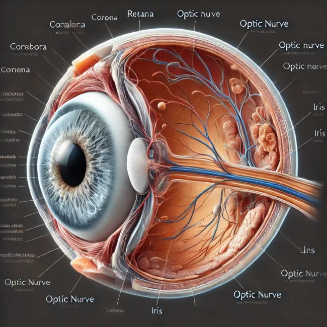
The human eye is an intricate organ responsible for converting light into visual information, enabling us to perceive our surroundings with precision. Every part of the eye—from the cornea and lens to the retina and optic nerve—plays a vital role in vision. This process involves retina light reception, which transforms light into electrical impulses, and optic nerve function, which transmits these signals to the brain for interpretation. Understanding eye anatomy terms is essential for grasping how vision works and diagnosing common eye conditions.
Vision begins when light enters through the pupil, the dark circular opening at the center of the iris, the coloured portion of the eye. The iris adjusts the pupil’s size to regulate the amount of light entering the eye. Behind the pupil lies the lens, a biconvex structure that fine-tunes focus, directing light toward the retina. This complex process, known as refraction, ensures that images are sharp and clear.
The Cornea and Lens: Controlling Focus and Light Entry
The cornea is the eye’s outermost transparent layer, functioning as the first point of contact for light. It plays a crucial role in refraction, bending incoming light to direct it toward the lens. This thin but durable layer protects the eye while enhancing focus. Behind the cornea is the anterior chamber, filled with aqueous humor, a clear fluid produced by the ciliary body that nourishes the eye and maintains internal pressure.

The lens, situated just behind the iris, adjusts shape to focus light on the retina. This process, known as accommodation, relies on the ciliary muscles, which control the lens’s curvature. If the lens fails to focus properly, conditions such as myopia (nearsightedness) or hyperopia (farsightedness) may develop, requiring corrective measures.
Retina and Photoreceptors: The Foundation of Vision
At the back of the eye, the retina light reception process converts light into electrical signals. The retina contains specialized photoreceptor cells: rods, which detect dim light and motion, and cones, which enable colour and detailed vision. The highest concentration of cones is found in the fovea centralis, a small depression in the macula responsible for sharp central vision.
The optic disk, located at the rear of the eye, is where the optic nerve function begins. This region, known as the blind spot, lacks photoreceptors, meaning it cannot detect light. However, the brain compensates for this absence, ensuring seamless vision. The optic chiasma, where optic nerves cross, allows the brain to merge visual input from both eyes, providing depth perception and a wide field of view.
The Supporting Layers: Choroid, Sclera, and Vitreous Humor
Beneath the retina, the choroid layer supplies blood and oxygen to the eye. This vascular layer prevents excessive light from scattering, enhancing visual clarity. The sclera, or “white of the eye,” provides structure and protection, reinforcing the eyeball’s shape.
The vitreous chamber, filled with vitreous humor, maintains eye pressure and helps transmit light to the retina. This gel-like substance stabilizes the eye’s structure, preventing retinal detachment and ensuring proper image formation.
Vision and the Science of Refraction
A fundamental process in human vision is refraction, the bending of light rays to achieve clear focus. The cornea and lens work together to direct light onto the retina, ensuring accurate image perception. However, imperfections in refraction can cause conditions such as astigmatism, requiring corrective lenses or surgery.
Examinations of the fundus of the eye—which includes the retina, optic disk, and macula—help detect conditions like macular degeneration and glaucoma. Regular eye exams are crucial for early diagnosis and treatment of vision-related disorders.
How the Eye Maintains Precision in Vision
The human eye’s ability to process images with accuracy depends on the seamless function of various components. From optic nerve function to retina light reception, each part plays a role in capturing, processing, and interpreting visual data. Understanding eye anatomy terms provides insights into common eye conditions and the importance of maintaining eye health.
Discover more from Business-News-Today.com
Subscribe to get the latest posts sent to your email.